

The sudden blindness that strikes days after a routine tooth extraction is like a nightmare no one sees coming. This real-life case, handled by vitreoretinal specialist Dr. Ashish Markan, involved a diabetic woman with unstable blood sugars who lost vision in one eye three days after extraction. Tests confirmed metastatic endophthalmitis, a wild infection where pus and bacteria overwhelm the inner chambers of the eye, threatening irreversible damage.
Understanding Metastasis endophthalmitis

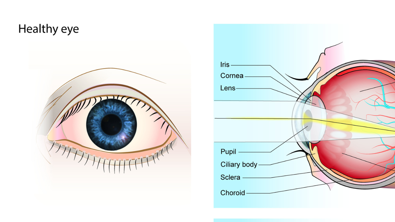
This condition strikes like lightning in the eye. Endophthalmitis means severe inflammation of the vitreous gel, the clear jelly that fills most of the eyeball—and the aqueous humor, the front fluid that nourishes the cornea and lens. “Metastatic” refers to bacteria that travel through the blood from a distant source, here the mouth during dental work. Tooth extractions cause transient bacteremia, a normal cycle where oral microbes such as strep or staphylococci slip into circulation. Most of the time, the body clears them within minutes. But when defenses falter, these bugs damage the eye, multiplying into pockets of pus that blur vision during the night. Early signs mimic pink eye, pain, redness, sensitivity to light that escalates to floaters and then blackout as the retinal cells starve.
Diabetes sets the vulnerable stage
Uncontrolled sugars increase the risks through no one’s fault. High glucose thickens the blood, damages white blood cells and feeds bacteria. Diabetics often have retinopathy scars, leaky vessels that invite invaders. Bacteremia from exports, common in 20-50 percent of operations, rarely spreads far, but compromised immunity changes that. Dr. Markan’s patient showed pus in eye fluids, confirming spread from the dental site to the eye via the bloodstream. The symptoms rapidly exploded, the vision falls on the movements of the hands, the hypopyon pus forms at the bottom like sediment in a storm.
Spotting it requires sharp eyes–Dr. Markan noticed sudden onset after the procedure, blurry insides, severe vision loss. Slit-lamp examinations reveal corneal edema, anterior chamber flare, vitreous opacities. B-scan ultrasound penetrates the clouds to map the pus pockets and the condition of the retina. Blood cultures hunt for the source of the germ, vitreous faucets become guilty for targeted drugs. Differentiate from sterile inflammation or uveitis, as it delays retinal scarring permanently. In this case, the clinical signs screamed infection, pus confirming the metastatic path.
Surgical battle for recovery
Urgency drove action. Dr. Markan moved to emergency vitrectomy, basic therapy, infected gel suction, eye wash, and intravitreal vancomycin infusion for Gram-positives and ceftazidime for Gram-negatives. Systemic antibiotics covered the spread throughout the body. Partial vision returned, a testament to the timing where the chances of full recovery range from 40-60 percent. They monitor for scarring, pressure spikes or detachment. Some need repeated shots, steroids tame the swelling. Success stories like hers highlight early intervention that reverses dire prognoses.
Because this condition requires respect
Endophthalmitis remains rare, with an impressive 0.1 percent post-dental spread, but is rapidly devastating. In the eyes there is no strong blood supply to resist, and the walls pus from the nutrients. Diabetics face 2-3 times higher odds, per incident series. Prevention weaves simple threads: strict sugar control before the procedure stabilizes the defense, good oral hygiene reduces the bacterial load. Regular diabetic eye screens catch vessels at risk. Awareness links the mouth to the health of the eyes, transient bugs that turn traitor only under the right storm. The doctor’s manipulation led to potential tragedy, highlighting how bodies are unexpectedly connected. A patient’s partial vision has returned to the spotlight, a condition where seconds count, proving that vigilance protects vision from hidden enemies.