Ian Shuman, DDS, AFAAID, MAGD
To meet daily demands of dental practices, a variety of impression and provisional techniques have been developed. An even greater number of materials have been developed to perform these techniques. The functions of these materials are very specific and depend on strength, durability, ability to capture detail and other properties.
Temporarily
Temporary crowns can be made chairside or in a laboratory. Temporary crowns and bridges made in the laboratory still require chair support, margin improvement and occlusal adjustment. To maximize accuracy and manage operating room time, an impression of the quadrant should be taken immediately after local anesthesia and before preparation. This pre-formed impression of the site simplifies temporary construction. This is a much more precise and effective method than acrylic “block-temps” – or pre-formed acrylic shells and stainless steel crowns. Acrylic has longer setting time, high shrinkage rate, bad taste and bad smell. Alternatively, cold-cured temporary composites create highly detailed, aesthetically pleasing temporaries extremely quickly and easily.
Regarding the impression material as a form for creating temporaries, it should provide enough stiffness to allow an accurate temporary to be formed, but be flexible enough to allow easy removal of the material set from this preliminary impression. Ideally, this is achieved by using a fast-setting, medium-body vinylpolysiloxane (VPS) impression material. Upon completion of the case and after a final impression, a composition-based temporary material is used. It should also provide a fast-setting elastic phase that allows easy removal from the prepared site (and midbody impression) and addition of additional material as needed. It should impart a high, natural gloss in a variety of shades without the need for polishing and only require removal of the air suspension layer, as with all composites. It should be durable and provide fracture toughness and high compressive strength to allow proper occlusion function.
Final appearance requirements
After the preparation of the tooth or teeth, the final impression is made. This can be achieved digitally via a scanner or, like the majority of professionals, using VPS impression material. Whether the VPS will be directly poured and modeled or exclusively scanned, this final impression must be absolutely accurate. The accurate impression is the primary source of information for the detailed reproduction of intraoral structures and a means of communication between the dentist and the dental technician. Any ambiguities in an impression will be transferred to the models and often go unnoticed until the final prosthesis is placed.
Precision dental restorations require a material with properties that support optimal impression taking from inception to casting. To maximize this accuracy, a highly hydrophilic VPS should be used. This feature allows excellent wetting of the prepared tooth and surrounding intraoral structures.
To make the actual impression-taking process highly efficient, the materials should have variable extraoral working times and short intraoral setting times. Also, an accurate impression must exhibit high hardness to ensure safe removal from the patient’s mouth. However, during intraoral removal, all impression materials will be stretched to varying degrees across the widest part of the teeth, changing their dimensions and potentially losing accuracy. This becomes moot provided the material exhibits excellent elastic recovery after removal. This guarantees the accuracy of the oral structures.1 In the laboratory, a VPS set with a high level of hydrophilicity—and thus accuracy—is maintained until pouring and/or scanning.
Final impression techniques
There are many final impression techniques that can be used depending on the clinical situation. However, for the majority of final crown and bridge impressions, a one- or two-step impression technique is used. Ideally, a family of impression materials with different viscosities and setting times would provide the greatest number of options, all depending on clinical needs.
Clinical case No. 1: Anterior crowns
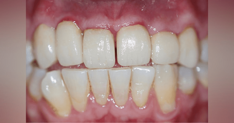
A 32-year-old man presented with recent dental treatment abroad. Six maxillary anterior teeth were individually crowned (figure 1).The patient’s chief complaint was severe pain in this area. He could not consume hot or cold food and drinks and could not use the teeth for cutting. A radiographic examination revealed grossly exaggerated crown contours, poorly fitted margins, and partially residual cementum (figure 2). Obstruction was evaluated and ruled out as the cause of the pain. After identifying the periapical radiolucency at the apex of the upper left lateral sector, it was suspected that the pulps of some or all of the crowned teeth were involved.
Figure 1: Presentation of a patient with six maxillary anterior teeth separately crowned

