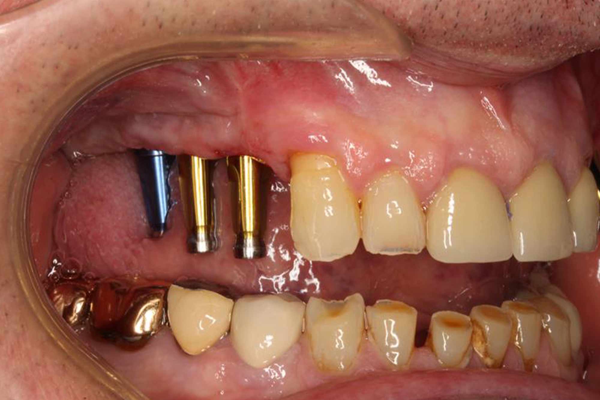
In the latest implant information, Nilesh Parmar explores the different methods of impression taking and the different materials available.
Impression techniques in dentistry remained unchanged for several years before the use of intraoral scanners.
Prior to this, impressions were made on conventional or special trays with a silicone compound usually the material of choice.
Over time the materials have been developed and perfected. Now they are easier to handle, set with improved hydrophilic properties.
Implant impressions require an impression to be inserted into the implant and a mold taken over it.
Two main methods of taking an implant impression are used today.

Close disk impressions
Here the clinician inserts an impression that faces a distinctive “head” on the implant.
A conventional silicone impression is taken (one-stage or two-stage techniques), with the impression remaining inside the implant when the mold is removed. The cover is then removed from the implant and inserted into the impression itself.
Various imprinting designs are available for this purpose. Many clinicians prefer this method because it is close to conventional crown and bridge impression techniques without requiring modification to the impression trays.
If the clinician places the implant deep below the bony crest, then less coping “head” is available above the gum line. This makes it difficult to firmly place the impression on the resulting material.
This technique has long been thought to be less accurate than open disc overlays. However, research would disagree with this statement.
Gallucci et al found that the closed disc method was as accurate as the open disc method for single unit restorations involving dental implants.
Open disk impressions
This technique is where the clinician screws an impression facing the implant with a long purchase screw. This screw should extend into and beyond the impression tray.
The impression tray will have holes in it so that the compensation screw can be accessed once the impression material has set.
The usual practice is to place wax over the hole made in the impression tray to allow the screw to enter. This will allow the clinician to loosen it once the material has set.
This technique is difficult to perfect at first. The fear is that the impression is locked in the mouth if access to the impression protection screw is not available. There is some thought that multiple implants, which require multiple holes in a stock impression disc, may lead to distortion in the final impression. Therefore, in these cases, clinicians prefer a special tray with impression funnels.
It is the method of choice for laboratory technicians and research shows it to be a superior impression method in multiple implant restorations compared to closed disc options (Papaspyridakos et al, 2014).
The open disc technique requires a rigid setting impression material, with Impregum (3M ESPE) being the common material to use.
Many patients do not like the chemical taste of this. The rigid set requires you to block the bottom pieces using wax or petroleum jelly to ensure you can remove the disc successfully.

Intraoral scanners
The open disc impression technique is the universal method of choice for implant impressions. However, we must not forget about the digital option.
The provision and development of intraoral scanners has dramatically changed the dental environment. The ability to digitally scan implant restorations and reconstructions has led to faster, less invasive and reproducible protocols for the fabrication of implant crowns and bridges.
There is a cost that goes with these methods. However, long-term savings are significant, combined with patient preference.
Conventional impression techniques still play a large role in implant impression techniques. But digital replacements are fast becoming commonplace.
Find out about previous implant information
Follow Dentistry.co.uk on Instagram to keep up with all the latest dental news and trends.
bibliographical references
Papaspyridakos P, Chen CJ, Gallucci GO, Doukoudakis A, Weber HP and Chronopoulos V (2014) Accuracy of implant impressions for patients with partial and complete endons: a systematic review. Int J Oral Implants Maxillofac 29(4): 836-45

